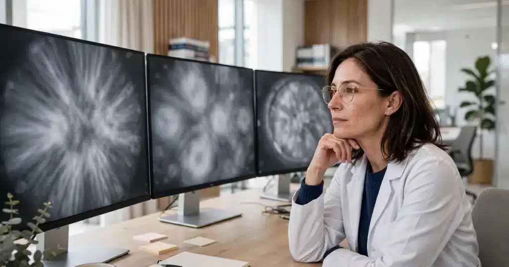
When a mammogram is read today, a specially trained doctor called a radiologist makes the decisions — but at many centres, computer software works alongside that radiologist, scanning the images, marking areas that may deserve a second look, and acting as a kind of tireless second reader. This is often described as “AI” (artificial intelligence), or by the older term computer-aided detection. The computer does not diagnose, does not replace the radiologist, and does not have the final say: it is a tool the human reader can use, question, or overrule. This article explains, in general terms, how that help is used and what it means for you.
Key Takeaways
- “AI” in mammogram reading means software that has been trained on large numbers of past images to recognise patterns worth a closer look.
- Radiologists use it in different ways: flagging areas on the image, acting as a second reader after their own first read, or helping them compare today’s images with your previous ones.
- The radiologist remains responsible for the result. A computer mark is a prompt to look again, not a finding of cancer.
- Being called back for more images does not mean a computer found something wrong — callbacks are a normal part of screening and most turn out to be nothing serious.
- How computer help is used varies between facilities, and some do not use it at all. Your imaging centre can tell you what happens where you are screened.
- Nothing about your part changes: attend screening as advised, and tell the team about any breast changes you have noticed, whatever any software says.
What “AI” Means in the Reading Room
A mammogram is a greyscale image full of overlapping tissue, and reading one well takes years of training. Computer-aided reading software is built by showing a program very large numbers of earlier mammograms — including images from people who were later found to have cancer and many who were not — until the program becomes good at noticing visual patterns that human readers consider worth attention: clusters of tiny bright specks, areas where the tissue pattern looks distorted, or a spot that has changed since a previous image.
The newer systems are usually described as AI because they learn patterns from examples rather than following fixed rules written by programmers. Either way, the important point for you is what the software produces: not a verdict, but marks. It highlights areas of the image and effectively says, “a human should look carefully here.” Our pillar guide to how breast cancer is found and diagnosed sets out the whole screening and diagnostic sequence, and computer help sits inside the reading step of that sequence.
The Ways Computer Help Is Used
Facilities use these tools in several ways, and a single centre may use more than one:
- Marking areas for a second look. As the radiologist reads, the software places small markers on areas its patterns flag. The radiologist looks at each marked area and decides whether it is normal tissue, needs more images, or needs further testing — and can dismiss a mark entirely.
- A second reader. In some services the software reads the images after (or before) the radiologist, in the same spirit as having a second radiologist double-check. Where two humans read each mammogram in some programmes, a computer may support or supplement one of those reads; arrangements differ by programme.
- Helping with comparisons. One of the most valuable things in reading a mammogram is comparing it with your earlier images, because change over time is a clue. Software can help align and present previous images alongside the new ones so differences stand out, as described in our article on why mammograms are compared with prior images.
- Supporting the workflow. Some centres use computer tools behind the scenes to organise worklists — for example, helping staff see which cases have already been read and which are waiting. This kind of use never touches your result directly.
If your screening leads to a closer look with another kind of imaging — for instance a targeted ultrasound prompted by something seen on an earlier scan — that follow-up is a separate, human-led step, described in our article on second-look ultrasound.

What the Computer Does Not Do
It helps to be equally clear about the limits. The software does not examine you, does not know your history or your symptoms, and does not decide whether you are called back, sent for a biopsy, or reassured. Those decisions rest with the radiologist and, where relevant, the wider care team, who can see the whole picture — the images, your previous studies, and the notes from your appointment. A mark from the software that the radiologist judges to be normal tissue goes no further.
It is also not a safety net that makes reading perfect. Human reading is not perfect either, which is why screening is a programme repeated over time rather than a single test, and why any new breast change between screenings should always be checked promptly rather than waiting for the next round. The difference between routine screening images and the more detailed imaging used when something needs investigating is explained in our comparison of diagnostic vs screening mammograms.
Practices also vary. Two centres in the same region may use different software, use the same software differently, or not use computer-aided reading at all. These tools are regulated as medical devices in the United States, but which tools a facility adopts, and how they fit its workflow, is a local decision. If you are curious about your own centre, asking is entirely reasonable — it is a normal question, and the answer tells you something about how your images will be read.
The Human Reader and the Computer, Side by Side
| The radiologist | The computer software | |
|---|---|---|
| What it works with | Your images, your previous studies, your clinical notes and reported symptoms | The images alone, compared against patterns learned from many past images |
| What it produces | The decision: normal, more images needed, or further tests — and your result letter | Marks and prompts suggesting areas worth a closer look |
| Can it overrule the other? | Yes — the radiologist can dismiss any computer mark | No — it cannot clear an area the radiologist is concerned about, and cannot order follow-up |
| Accountability | The radiologist and the screening programme are responsible for your result | None — it is a tool, used under the programme’s oversight |
Reading the table, the pattern is simple: the software is good at tireless pattern-spotting across a large image, and the human is good at judgement — weighing history, change, and context. Screening programmes that use computer help are trying to combine the two strengths, not to hand the job over.

Questions to Ask
- (Your imaging centre) Does this centre use computer-aided or AI software when mammograms are read?
- (Your imaging centre) Who makes the final decision on my result — and will a person always read my images?
- (Your doctor) I was called back for more images. Can you explain what the radiologist wants to look at more closely?
- (Your doctor) My result mentions marks or areas of interest. What happens next, and how soon?
- (Your care team) If I notice a breast change between screenings, what should I do — wait for the next mammogram or contact you?
Frequently Asked Questions
Will a computer decide my mammogram result?
No. Where computer help is used, it supports the radiologist by marking areas to review. The radiologist reads your images, weighs your history and previous studies, and makes the decision behind your result. If your centre uses such software, it works under the screening programme’s oversight, and a person is always responsible for the outcome.
Does AI make mammograms foolproof?
No test is foolproof, with or without computer help. Mammograms can miss some cancers and can flag areas that turn out to be harmless — that is true of human reading too. This is why screening is repeated at intervals, why comparison with your previous images matters, and why a new breast change should always be reported promptly rather than waiting for the next screening round.
I was called back after my mammogram. Does that mean the computer found cancer?
No. A callback means the radiologist wants a clearer look at one area — often with different mammogram views or an ultrasound. Most callbacks end in reassurance: overlapping tissue, a harmless cyst, or an area that looks normal on the closer images. The callback decision is made by the radiologist, not by software, and it is a routine part of how screening works.
Can I ask whether my centre uses AI?
Yes. It is a fair and increasingly common question. The centre can tell you whether computer-aided reading is part of its process and how the final result is reached. There is no requirement for a centre to use it, and not using it does not mean your images are read carelessly — many excellent services rely on one or two human readers alone.
Does computer help change anything I need to do?
Nothing at all. Attend screening when you are invited, keep appointments for any follow-up imaging, and mention any breast changes or symptoms you have noticed when you attend — the human team needs that information, and no software can supply it. If you are unsure what your result letter means, your doctor or the screening centre can go through it with you.
Related Articles
- How Breast Cancer Is Found and Diagnosed: The Complete Guide
- 2D vs 3D Mammogram: How They Compare
- Why Mammograms Are Compared With Your Prior Images
- Diagnostic vs Screening Mammogram: What Is the Difference?
- Second-Look Ultrasound: Rechecking an MRI Finding With Ultrasound
This article is for general educational purposes only and is not medical, legal, or financial advice. It does not replace professional care. Talk to your doctor or care team about your own situation. If you think you may have a medical emergency, call 911.