
After breast cancer treatment ends, follow-up monitoring usually rests on three quiet pillars — regular check-in appointments, physical examinations, and mammograms of any remaining breast tissue — rather than on routine whole-body scans for people who feel well. That surprises many survivors, who expect scans to be the main safety net. In fact, scans have real limits as well as real uses: they are excellent at answering a specific question, but as a general early-warning system in someone who feels well, they can miss small changes, raise false alarms, and find things that turn out to be harmless. This guide explains what monitoring imaging can and cannot show, and why your own care team — not any standard timetable — sets your follow-up plan.
Key Takeaways
- Follow-up after treatment is usually built on scheduled check-ins, physical examination, scheduled mammograms, and you reporting new symptoms — not on routine whole-body scanning.
- Scans such as CT, PET, and bone scans are powerful tools for answering a specific question — a symptom, a finding, a decision — but they are not a reliable net for catching every possible change early in someone who feels well.
- Monitoring imaging has built-in limits: very small changes may not show, results can be ambiguous, and scans commonly turn up incidental findings that need follow-up but prove harmless.
- A scan result is a snapshot of what imaging could detect on that day, interpreted alongside your history and examination — reassuring, but never a guarantee about the future.
- There is no single monitoring schedule this article can give you: your plan is set by your own care team, based on your treatment and situation, and it is always reasonable to ask them to explain it.
How Follow-Up Monitoring Usually Works
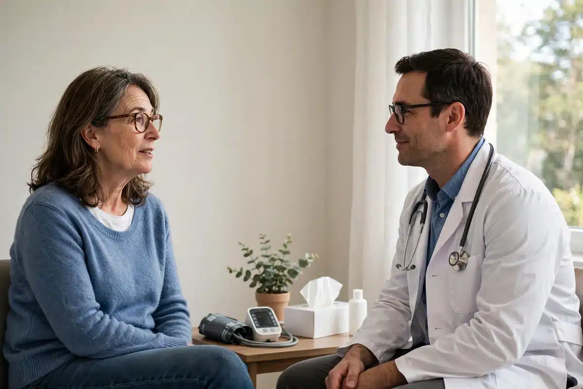
When treatment ends, care changes gear rather than stopping. In most follow-up plans, the surveillance work is shared between the clinic and you: scheduled visits where your team asks how you are and examines you; mammograms, which are X-rays of the breast, taken of any remaining breast tissue on the plan your team sets; and you, knowing your own normal and reporting changes between appointments. Our pillar guide to life after breast cancer and survivorship lays out this whole phase, and our article on the rhythm of follow-up visits describes how appointments usually settle over time.
Newer survivors sometimes assume this is a “light” version of monitoring, and that more scanning would automatically mean safer. The teams who design follow-up plans think about it differently: a test earns its place when it can find something early enough, and clearly enough, to change what happens next — without creating more harm through false alarms and unnecessary procedures than benefit. Whole-body scans in people who feel well have historically struggled to meet that bar, which is why they are usually held for when there is a specific question to answer. General background on follow-up care is published by the National Cancer Institute at cancer.gov.
What Scans Can Show
None of this means scans are weak tools — used at the right moment, they are remarkable. Depending on the type, monitoring imaging can:
- Show the size, shape, and position of a specific finding — a lump, a thickened area, a change seen on a mammogram — precisely enough for the team to decide what to do about it.
- Compare like with like over time. A current image set against your previous ones can show whether an area is stable, shrinking, or growing, which is often more informative than any single picture.
- Look at a particular area of concern — bones, chest, abdomen — when a symptom or examination finding points there.
- Guide a procedure, such as placing a needle accurately to sample tissue from a specific spot.
- Check how treatment is working in situations where ongoing treatment is being monitored, where change over time is the whole question.
Mammography deserves its own mention because it is the one imaging test that usually is scheduled as monitoring: it is designed for breast tissue, it is compared against your prior mammograms, and changes in the breast — including in the treated breast, where scar tissue makes the picture more complex — are exactly what it is built to flag. Our explainer on what happens during a mammogram covers the appointment itself.
What Scans Cannot Show
The limits are just as important to understand, because unrealistic expectations of imaging cause a great deal of avoidable distress:
| Expectation | The reality |
|---|---|
| “A clear scan means there is definitely no cancer anywhere” | A scan shows what was detectable, in the areas imaged, at the resolution of that technology, on that day. Very small clusters of cells are below what any scan can resolve — which is one reason follow-up continues even after reassuring imaging. |
| “A scan can tell whether a spot is cancer” | Imaging shows appearances, not identities. A suspicious-looking area may be scar tissue, inflammation, or a benign change; a quiet-looking situation can still hold a question. Tissue sampling is what identifies. |
| “Scanning more often catches everything earlier” | More frequent scanning mainly finds more ambiguous things sooner. Each ambiguity can trigger more imaging, waiting, and sometimes biopsies of things that were never dangerous. |
| “A scan replaces examination and symptoms” | A physical examination and your own report of a new change can flag things a scheduled scan would not be looking for, and can trigger targeted imaging at the moment it is actually needed. |
Put plainly: monitoring imaging is a set of excellent spotlights, not a floodlight over the whole field at all times. That is not a failure of the technology — it is the honest shape of what pictures of the body’s insides can do.
False Alarms and Incidental Findings
Two related experiences catch many people off guard. The first is the callback: a mammogram or scan shows something the radiologist cannot confidently call harmless, so more images, a closer look, or a biopsy follow — and the eventual answer is benign. Treated breasts, with their scar tissue and post-treatment changes, generate these situations readily, and our guide to abnormal mammogram results walks through how callbacks usually unfold.
The second is the incidental finding: a scan done for one reason spots something unrelated elsewhere — a spot on a lung, a cyst on the liver, a thyroid nodule. Most such findings are harmless features of an ordinary body, but each one has to be assessed, which can mean more appointments and more waiting in a season of life when your tolerance for medical uncertainty is already spent. Neither experience means anyone made a mistake. Both are the predictable cost of looking — and they form part of why follow-up plans use blanket scanning cautiously, reserving it for when the likely benefit outweighs this certain burden.
When Scans Are Used in Follow-Up
Scans absolutely have a place after treatment — a targeted one. In follow-up, imaging beyond mammograms is typically brought in when:
- You or your team find a new change — a lump, thickening, skin change, or anything that feels different and does not settle.
- A new or persistent symptom needs explaining — for example, ongoing pain in one place, or a symptom your team wants to look into rather than watch.
- A mammogram or examination shows something that needs a closer, different kind of look.
- Your individual situation — the details of your original cancer and treatment — leads your team to build imaging into your particular plan.
Notice what this list is: imaging answering questions as they arise, used promptly when there is something to answer. That is a different thing from scanning on a fixed drumbeat regardless — and it means a new symptom from you can set imaging in motion faster than any schedule would. Which plan applies to you, and what imaging it includes, is exactly the conversation to have with your own team; plans genuinely differ, and a fellow survivor’s plan looking different from yours is not evidence that either is wrong.

Your Part of the System
Because monitoring leans on examination and reporting, you are not a passive passenger in it — you are one of its instruments. The useful habits are simple: keep the follow-up appointments on the plan your team sets out (it should be written into your survivorship care plan, alongside your treatment summary); stay familiar with how your body normally looks and feels now, including the treated side, whose normal has changed; and report new or persistent changes when you notice them rather than saving them up for the next scheduled visit.
It is also worth naming the emotional weather around all of this. Waiting for any scan or result can stir up the fear of recurrence that most survivors know well, and that fear can make a modest monitoring plan feel inadequate even when it is carefully designed. If that feeling is strong, say so at your appointment. Understanding the reasoning behind your own plan — which is a fair and ordinary thing to ask for — is often more settling than any single test.
Questions to Ask Your Doctor
- What does my follow-up monitoring plan consist of, and can you write it down for me?
- Which imaging is part of my plan, and which is held in reserve in case something comes up?
- Why is routine whole-body scanning not part of my plan — can you explain the reasoning for my situation?
- What changes or symptoms should make me contact you straight away rather than waiting?
- If I needed a scan, what would it be looking for, and what would happen after each possible result?
- Who reviews my mammograms, and how will I receive the results?
Frequently Asked Questions
Why won’t my team just scan me regularly so we catch anything early?
Because for someone who feels well, routine whole-body scanning has not proven to be the reliable early-warning system it sounds like: it can miss very small changes, it frequently raises alarms over harmless findings, and each alarm can lead to more tests and procedures. Follow-up is instead built on examination, mammograms, and prompt targeted imaging when something specific needs answering — a design your team can explain for your own case.
If a scan can’t rule everything out, what is the point of follow-up?
Follow-up layers several kinds of watching, each covering the others’ gaps: examination by someone who knows your history, mammograms compared against your previous images, your own knowledge of your body’s normal, and scans deployed quickly when there is a question. No single layer is perfect; the combination is the safety net.
My friend’s follow-up includes scans and mine doesn’t. Should I worry?
Not on that evidence alone. Plans are individual — shaped by the type and stage of the original cancer, the treatment given, and each team’s judgement. If the difference troubles you, ask your own team what sits behind your plan. That conversation is routine, and a good team expects it.
What is an incidental finding, and what happens if I get one?
It is an unexpected spot or feature found by a scan that was looking at something else. Most turn out to be harmless variations of a normal body. Your team will assess it — sometimes by simply noting it, sometimes with a follow-up image or test — and will explain what they found and why they are or are not concerned.
Should I ask for a scan if I feel anxious but have no symptoms?
Tell your team about the anxiety itself — that is a legitimate and common part of survivorship, and they hear it often. Sometimes a conversation about your plan, your actual situation, and what would trigger imaging is more relieving than a scan would be, because a scan’s reassurance tends to fade while its ambiguities can linger. If you and your team decide together that imaging would help, that is a shared clinical decision, made properly.
Related Articles
- Life After Breast Cancer: The Complete Survivorship and Follow-Up Guide
- Follow-Up Visits After Breast Cancer: How the Rhythm of Care Usually Works
- Follow-Up Care Plans When Treatment Ends
- Fear of Recurrence: Coping After Treatment
- What Is a Survivorship Care Plan? What It Includes and How to Get Yours
This article is for general educational purposes only and is not medical, legal, or financial advice. It does not replace professional care. Talk to your doctor or care team about your own situation. If you think you may have a medical emergency, call 911.